Jean Baptiste Joseph Fourier (1768-1830) a French mathematician and physicist developed the mathematical methods for converting data between the time domain and the frequency domain – a method now used in a critical step in utilizing NMR and MRI data.
In 1938 Isidor Isaac Rabi, physics professor at Columbia University, sent a beam of molecules through a magnetic field and demonstrated that they could be made to emit radio waves at specific frequencies.
Magnetic Resonance Imaging was broadly introduced to the scientific community in 1973, when Paul C. Lauterbur published a technique to locate the positions of water molecules within the body, which in turn could provide the basis of images of specific cross sections.
Soon thereafter, Peter Mansfield at the University of Nottingham introduced critical methods for efficient image generation, including slice selection and fast “snapshot” acquisition schemes wherein entire 2-D images could be obtained in a few tens of milliseconds.
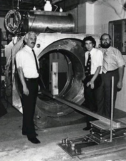
Dr Raymond Damadian, a professor at the SUNY Downstate Medical Center, was the first to attempt a magnetic resonance scan of a human body. The first MRI body scan was conducted on a human on July 3, 1977. His experiment helped develop the medical technology now known as the MRI scanner.
In 1980 saw the introduction of the first commercial MR imaging system, based on Damadian’s field focused nuclear MR technique. The early 1980s saw the first publications pertaining to the brain, spine, chest, breast, abdomen, and pelvis.
With the invention of the Fourier transform–based spinwarp imaging technique in 1980, it became possible to acquire a two-dimensional image within a reasonable imaging time.
History of MRI Scan
The History and Evolution of Soy Sauce Process
-
Soy sauce, an indispensable condiment in Asian cuisine, has a history
spanning over 2,500 years. Originating in ancient China, its development
reflects cul...


